
Meibography
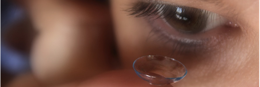
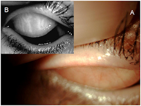 Meibomian gland dysfunction (MGD) is one of the most common abnormalities in ophthalmic practice causing an abnormality of the tear film lipid layer resulting in the evaporative dry eye. Meibography is the only in-vivo means to evaluate the morphology of the meibomian glands. One principal is the transillumination of the everted lid the other one the direct illumination, named the non-contact meibography. In transillumination the lid is everted over a light source while non-contact meibography consist of a slit lamp equipped with an infrared charge-coupled device video camera and an infrared transmitting filter to allow the observation of the everted lid without contact to the instrument (Arita et al 2008).
 To be independent of a slit lamp microscope, we were first describing the benefit of an IR-CCD camera with near-optic in non-contact meibography (portable non-contact meibograph (PNCM)) or the built-in IR cameras of common ophthalmic instruments to be designed for pupillometry.
Furthermore we were first having developed computerized analyses of the meibomian gland drop-out aand are continuously investigating in this issue.
The concept is on the market now.
The multi-functional instruments "EyeTop topographer", "Sirius Scheimpflugkamera" and "Cobra Fundus Camera" of b o n Optic, Lübeck, Germany are now equipped with that technology using their built-in IR cameras.
We feel that this will really push research and clinical practice in MGD.
